Myocarditis Not Recovered in 80% at 6 Months after Vaccination
Worrisome Serial MRI Results in Adolescents after Primary mRNA Series
By Peter A. McCullough, MD, MPH
Every cardiology office in America should be recognizing COVID-19 vaccine-induced myocarditis presenting in young persons, 90% are male, with chest pain, effort intolerance, arrhythmias, and cardiac arrest after injections of mRNA vaccines. As I see these patients, the common question is “when is this over?”. While ECG and blood tests tend to normalize quickly, my concern is that ongoing inflammation is occurring due to continued production of Wuhan Spike protein coded by the long lasting Pfizer or Moderna mRNA vaccines. While blood tests can give inferences on inflammation, cardiologists also use cardiac magnetic resonance imaging (MRI) to visualize the inflammation, establish the diagnosis, and craft a prognosis. We would hope young teenagers would resolve their MRI results and go on with life. A recent report to the contrary caught my attention.
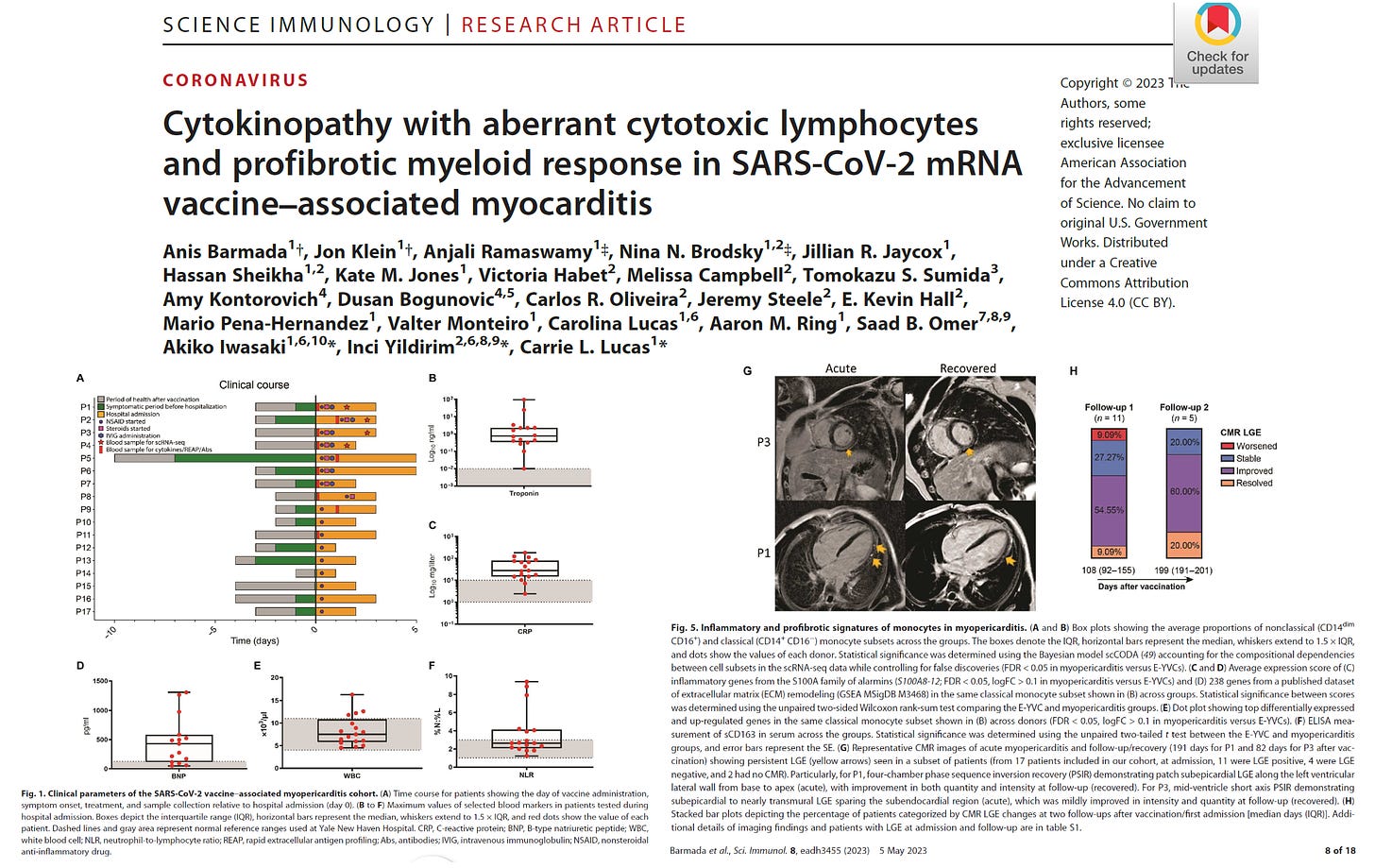
Barmada et al studied a clinical cohort consists of 23 patients hospitalized for vaccine-associated myocarditis and/or pericarditis. The cohort was predominately male (87%) with an average age of 16.9 ± 2.2 years (ranging from 13 to 21 years). Patients had largely noncontributory past medical histories and were generally healthy before vaccination. Most patients had symptom onset 1 to 4 days after the second dose of the BNT162b2 mRNA vaccine. Six patients either first experienced symptoms after a delay of >7 days after vaccination or were incidentally positive for SARS-CoV-2 by polymerase chain reaction (PCR) testing upon hospital admission; these six patients were thus excluded from further analyses, although they potentially reflect the breadth of clinical presentations of vaccine-associated myopericarditis. The remaining cohort of 17 patients showed no evidence of recent prior SARS-CoV-2 infection, with antibodies to spike (S) protein but not to nucleocapsid (N) protein and negative nasopharyngeal swab reverse transcription quantitative PCR at hospital admission.

While the authors clearly show high levels of inflammatory markers, my attention was drawn to the follow-up MRI scans. As shown in the figure, only 20% had resolved their abnormalities (late gadolinium enhancement) at over six months (199 days). This paper raises questions: 1) is there ongoing heart damage and inflammation at six months? 2) does the LGE in 80% represent a permanent “scar” putting these children at risk for future cardiac arrest? These data strongly call for large scale research into this emerging problem given the large number of potential young persons at risk.
If you find “Courageous Discourse” enjoyable and useful to your endeavors, please subscribe as a paying or founder member to support our efforts in helping you engage in these discussions with family, friends, and your extended circles.


I guess this is what the CDC calls "mild" myocarditis?
Since damage to heart muscle tissues due to myocarditis is effectively irreversible, one wonders how the 20% in the cohort were shown to have resolved on later MRI imaging? That the 80% did not resolve is beyond troubling, and for these young people it is life-altering, follow up data are needed though the prognosis is not good unfortunately.